Research Interests
Topics: Neuromotor Control, Neurodegenerative Diseases, mild Traumatic Brain Injury, Tumor Imaging
Approaches: Brain Connectome Analysis, Tractography, Radiomics, Machine Learning
Modalities: Diffusion Tensor Imaging, functional Magnetic Resonance Imaging (fMRI), functional Near Infrared Spectroscopy (fNIRS), Electroencephalography (EEG)
Principal Investigator: Chia-Feng Lu
盧家鋒, Ph.D. (alvin4016@ym.edu.tw)
Please Click Here for Full English CV and Publication List.
舊版中文研究介紹 Click Here

Summary of Works @ 2018.5.7
1. Machine Learning-Based Radiomics for Molecular Subtyping of Gliomas, < MRP Platform & Models >
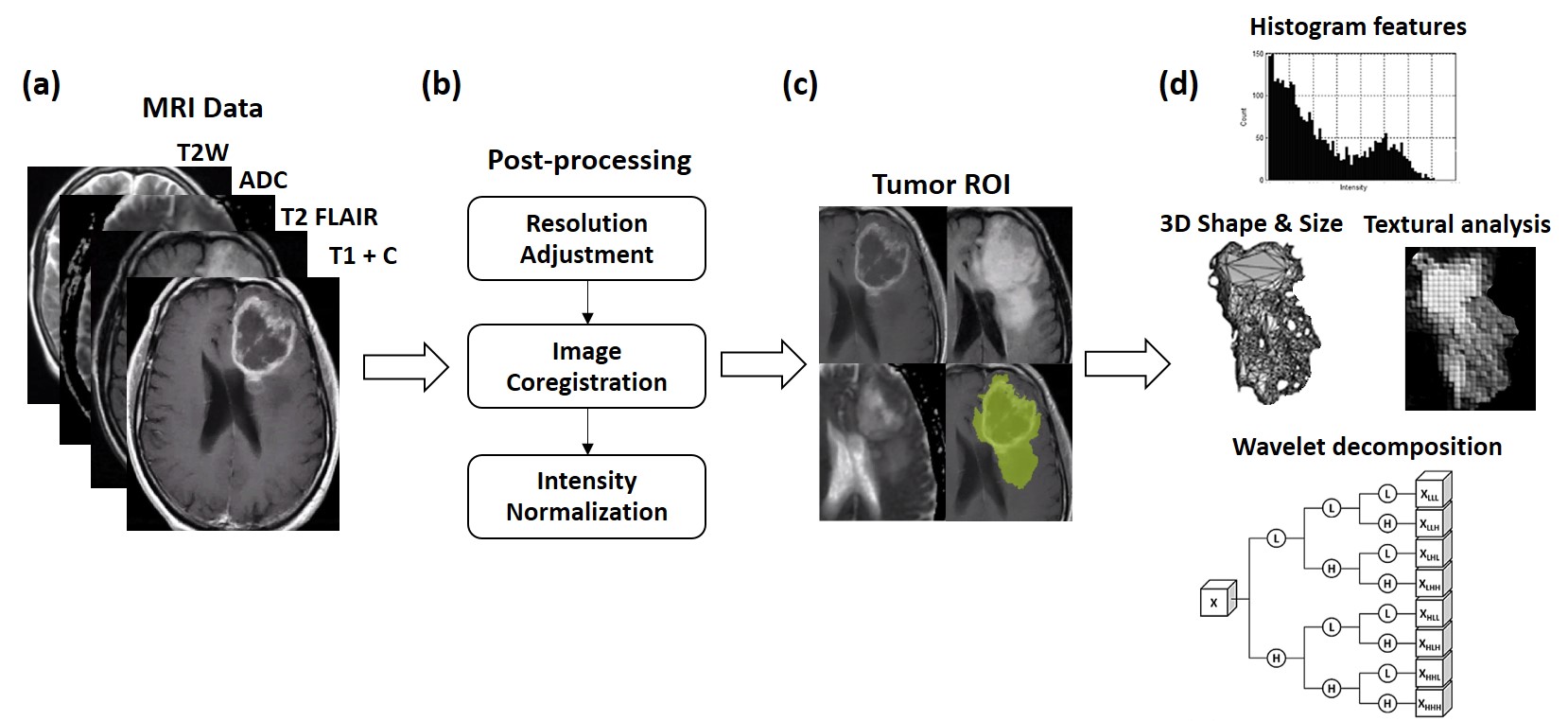
Purpose: The new classification announced by the World Health Organization in 2016 recognized five molecular subtypes of diffuse gliomas based on isocitrate dehydrogenase (IDH) and 1p/19q genotypes in addition to histological phenotypes. We aim to determine whether clinical magnetic resonance imaging (MRI) can stratify these molecular subtypes to benefit the diagnosis and monitoring of gliomas.
Experimental Design: The data from 456 subjects with gliomas were obtained from The Cancer Imaging Archive. Overall, 214 subjects, including 106 cases of glioblastomas and 108 cases of lower-grade gliomas with preoperative MRI, survival data, histology, IDH, and 1p/19q status were included. We proposed a three-level machine-learning model based on multimodal MR radiomics to classify glioma subtypes. An independent dataset with 70 glioma subjects was further collected to verify the model performance.
Results: The IDH and 1p/19q status of gliomas can be classified by radiomics and machine-learning approaches, with areas under receiver operating characteristic curves between 0.922 and 0.975 and accuracies between 87.7 and 96.1% estimated on the training dataset. The test on the validation dataset showed a comparable model performance with that on the training dataset, suggesting the efficacy of the trained classifiers. The classification of 5 molecular subtypes solely based on the MR phenotypes achieved a 81.8% accuracy, and a higher accuracy of 89.2% could be achieved if the histology diagnosis is available.
Conclusion: The MR radiomics-based method provides a reliable alternative to determine the histology and molecular subtypes of gliomas.
Relevant Publications
(1) Chia-Feng Lu, Fei-Ting Hsu, Kevin Li-Chun Hsieh, Yu-Chieh Jill Kao, Sho-Jen Cheng, Bo-Kai Hsu, Ping-Huei Tsai, Ray-Jade Chen, Chao-Ching Huang, Yun Yen, Cheng-Yu Chen. Machine Learning-Based Radiomics for Molecular Subtyping of Gliomas. Clinical Cancer Research, accepted, 2018. (SCI)
(2) Chia-Feng Lu, Fei-Ting Hsu, Li-Chun Hsieh, Yu-Chieh Jill Kao, Cheng-Yu Chen. Machine Learning based Classification of Glioma Subtypes Characterized by MR Radiomics. The 103rd Annual Meeting of Radiological Society of North America, Scientific Posters, Chicago, USA, Nov 26-Dec 1, 2017.
(3) Cheng-Yu Chen, Chia-Feng Lu, Fei-Ting Hsu, Paul Blakeley, Li-Chun Hsieh, Yu-Chieh Jill Kao, Ping-Huei Tsai, Yung-Hsiao Chiang, Wan-Yuo Guo, Ming-Hsong Chen, Chih-Chun Wu, Liang-Wei Chen, Hung Wen Kao, Yuan-Hao Chen, Hsin-I Ma. Personalized Survival Prediction Using Random Forest Survival Model on MR Radiomic Features in Gliomas. The 103rd Annual Meeting of Radiological Society of North America, Scientific Papers (Oral), Chicago, USA, Nov 26-Dec 1, 2017.
(4) Chia-Feng Lu, Fei-Ting Hsu, Li-Chun Hsieh, Yu-Chieh Jill Kao, Hua-Shan Liu, Ping-Huei Tsai, Pen-Yuan Liao, Cheng-Yu Chen. Imaging Angiogenesis Genotype of Glioblastoma by Radiomic Features of Multimodality MRI. The 24th Annual Meeting & Exhibition of ISMRM, Singapore, May 7-13, 2016.
2. Disrupted Cerebellar Connectivity Reduces Whole-Brain Network Efficiency in Multiple System Atrophy
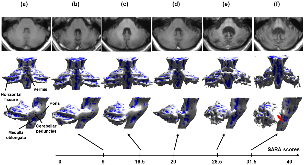
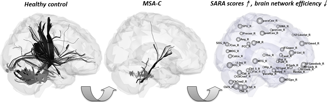
Purpose: Multiple system atrophy of the cerebellar type is a sporadic neurodegenerative disorder of the central nervous system. We hypothesized that the white matter degeneration of the cerebellum and pons in this disease may cause a breakdown of cerebellar
structural networks and further reduce the network efficiency of cerebellar-connected cerebral regions.Materials and Methods: Diffusion tensor tractography was used to construct the structural networks of 19 cerebellar-type multiple
system atrophy patients, who were compared with 19 age- and sex-matched controls. Graph theory was used to assess the small-world properties and topological organization of structure networks in both the control and patient groups.Results: Our results showed that the cerebellar- type multiple system atrophy patients exhibited altered small-world architecture with significantly increased characteristic shortest path lengths and decreased clustering coefficients. We also found that white matter degeneration in the cerebellum was characterized by reductions in network strength (number and integrity of fiber connections) of the cerebellar regions, which further induced extensively decreased network efficiency for numerous cerebral regions. Finally, we found that the reductions in nodal efficiency of the cerebellar lobules and bilateral sensorimotor, prefrontal,
and basal ganglia regions negatively correlated with the severity of ataxia for the cerebellar-type multiple system atrophy patients.Conclusion: This study demonstrates for the first time that the brains of cerebellar-type multiple system atrophy patients exhibit disrupted topological organization of white matter structural networks. Thus, this study provides structural evidence of the
relationship between abnormalities of white matter integrity and network efficiency that occurs in cerebellar- type multiple system atrophy.
Relevant Publications
(1) Chia-Feng Lu, Po-Shan Wang, Yuan-Lin Lao, Hsiu-Mei Wu, Bing-Wen Soong, Yu-Te Wu. Medullo-ponto-cerebellar White Matter Degeneration Altered Brain Network Organization and Cortical Morphology in Multiple System Atrophy. Brain Structure & Function, 219(3):947-958, 2014. (SCI)
(2) Chia-Feng Lu, Bing-Wen Soong, Hsiu-Mei Wu, Shin Teng, Po-Shan Wang, Yu-Te Wu. Disrupted Cerebellar Connectivity Reduces Whole-Brain Network Efficiency in Multiple System Atrophy. Movement Disorders, 28(3):362-369, 2013. (SCI)
(3) Po-Shan Wang, Chien-Li Yeh, Chia-Feng Lu, Hsiu-Mei Wu, Bing-Wen Soong, and Yu-Te Wu. The involvement of supratentorial white matter in multiple system atrophy: a diffusion tensor imaging tractography study. Acta Neurologica Belgica 2016: 1-8. (SCI)
3. Reorganization of functional connectivity during the motor task using EEG time–frequency cross mutual information analysis
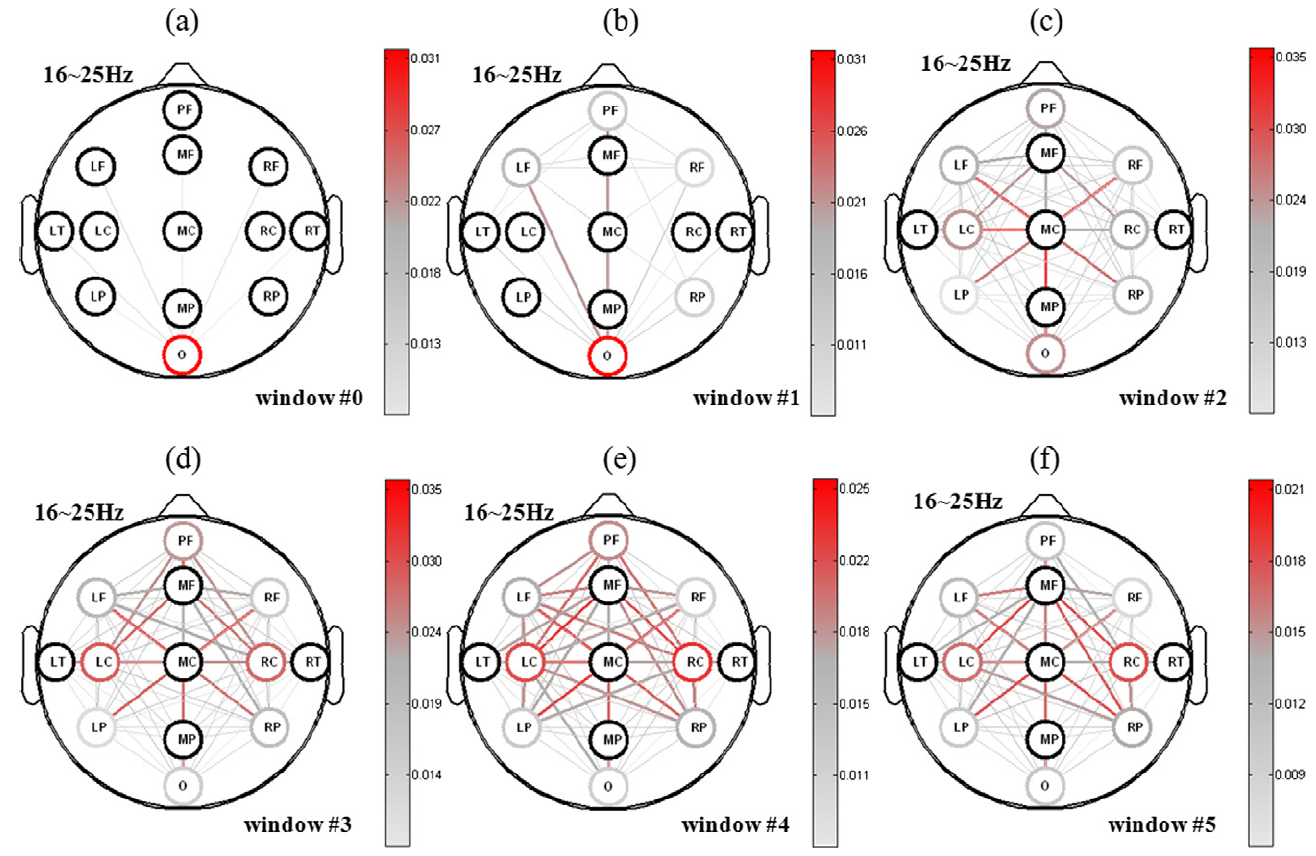
Objective: This study investigates the functional organization of cortical networks during self-determinant arm movement using the time sequences of the alpha (8–12 Hz) and beta (16–25 Hz) bands.
Methods: The time–frequency cross mutual information (TFCMI) method was used to estimate the EEG functional connectivity in the alpha and beta bands for seven healthy subjects during four functional states: the resting, preparing, movement-onset, and movement-offset states.
Results: In the preparing state, the maintenance of the central-executive network (CEN, prefrontal–parietal connection) suppressed the motor network in the alpha band to plan the next movement, whereas the CEN was deactivated in the beta band to retain visual attention (the frontal–occipital connection). A significant decrease of the CEN in the alpha band occurred after a visual cue in the movement-onset state, followed by a significant increase in motor-network connectivity in the beta band until the movement-offset state.
Conclusion: The temporal-spectral modulation mechanism allows the brain to manifest multiple functions subject to energy budget.
Significance: The TFCMI method was employed to estimate EEG functional connectivity and effectively demonstrate the reorganization process between four functional states.
Relevant Publications
(1) Chia-Feng Lu, Shin Teng, Chih-I Hung, Po-Jung Tseng, Liang-Ta Lin, Po-Lei Lee, Yu-Te Wu. Reorganization of Functional Connectivity during the Motor Task Using EEG Time-frequency Cross Mutual Information Analysis. Clinical Neurophysiology, 122:1569-1579, 2011. (SCI)
(2) Chia-Feng Lu, Chih-I Hung, Po-Jung Tseng, Liang-Ta Lin, Zun-Yun Wang, Yu-Te Wu. Recognition of Resting and Movement-Related Electroencephalography (EEG) Using Time-Frequency Cross Mutual Information. In Proceedings of CACS International Automatic Control Conference, Taipei, Taiwan, November 27-29, 2009.
(3) Chia-Feng Lu, Chih-I Hung, Po-Jung Tseng, Liang-Ta Lin, Zun-Yun Wang, Yu-Te Wu. Recognition of Arm-Movement Electroencephalography (EEG) Using Motor-Related Intrinsic Mode Functions Filtering and Cross Mutual Information. World Congress, Munich, Germany, 2009.
4. Functional MRI Studies of Psychiatric Disorders and Motor Control
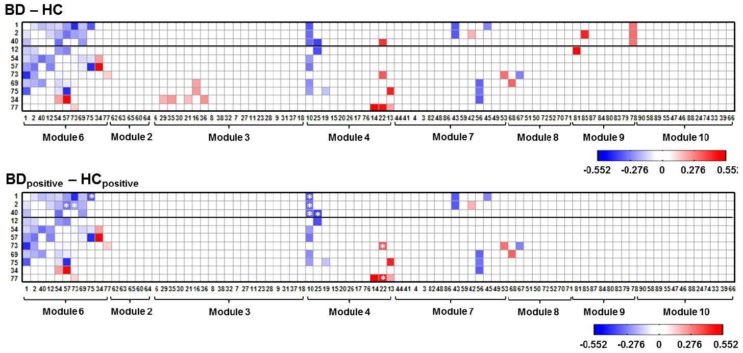
Purpose: Bipolar disorder is characterized by internally affective fluctuations. The abnormality of inherently mental state can be assessed using resting-state fMRI data without producing task-induced biases. In this study, we hypothesized that the resting-state connectivity related to the frontal, striatal, and thalamic regions, which were associated with mood regulations and cognitive functions, can be altered for bipolar disorder.
Methods: We used the Pearson’s correlation coefficients to estimate functional connectivity followed by the hierarchical modular analysis to categorize the resting-state functional regions of interest (ROIs). The selected functional connectivities associated with the striatal-thalamic circuit and default mode network (DMN) were compared between bipolar patients and healthy controls.
Results: Significantly decreased connectivity in the striatalthalamic circuit and between the striatal regions and the middle and posterior cingulate cortex was observed in the bipolar patients. We also observed that the bipolar patients exhibited significantly increased connectivity between the thalamic regions and the parahippocampus. No significant changes of connectivity related to the frontal regions in the DMN were observed.
Conclusion: The changed resting-state connectivity related to the striatal-thalamic circuit might be an inherent basis for the altered emotional and cognitive processing in the bipolar patients.
Relevant Publications
(1) Shin Teng#, Chia-Feng Lu#, Po-Shan Wang, Cheng-Ta Li, Pei-Chi Tu, Chih-I Hung, Tung-Ping Su, Yu-Te Wu. Altered Resting-State Functional Connectivity of Striatal-Thalamic Circuit in Bipolar Disorder. PLoS ONE, 9(5): e96422, 2014. (SCI) #: equal contribution.
(2) Yuan-Lin Liao, Po-Shan Wang, Chia-Feng Lu, Chih-I Hung, Cheng-Ta Li, Ching-Po Lin, Jen-Chuen Hsieh, Tung-Ping Su, Yu-Te Wu. Cortical Shape and Curvedness Analysis of Structural Deficits in Remitting and Non-remitting Depression. PLoS ONE, 8(7):e68625, 2013. (SCI)
(3) Shin-Yi Chiou, Ray-Yau Wang, Kwong-Kum Liao, Yu-Te Wu, Chia-Feng Lu, Yea-Ru Yang. Co-activation of primary motor cortex ipsilateral to muscles contracting in a unilateral motor task. Clinical Neurophysiology, 124:1353-1363, 2013. (SCI)
(4) Shin-Yi Chiou, Ray-Yau Wang, R. Edward Roberts, Yu-Te Wu, Chia-Feng Lu, Kwong-Kum Liao, Yea-Ru Yang. Fractional Anisotropy in Corpus Callosum is Associated with Facilitation of Motor Representation during Ipsilateral Hand Movements. PLoS ONE, 9(8):e104218, 2014. (SCI)
5. Maintaining Gait Performance by Cortical Activation during Dual-Task Interference: A Functional Near-Infrared Spectroscopy Study
Background: In daily life, mobility requires walking while performing a cognitive or upper-extremity motor task. Although previous studies have evaluated the effects of dual tasks on gait performance, few studies have evaluated cortical activation and its association with gait disturbance during dual tasks.
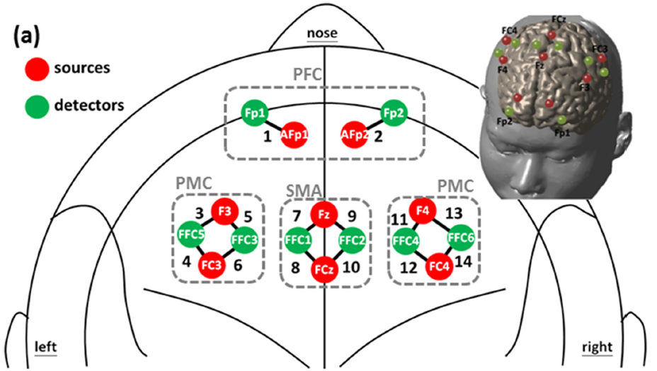
Materials & Methods: In this study, we simultaneously assessed gait performance and cerebral oxygenation in the bilateral prefrontal cortices (PFC), premotor cortices (PMC), and supplemental motor areas (SMA), using functional near-infrared spectroscopy, in 17 young adults performing dual tasks. Each participant was evaluated while performing normal- pace walking (NW), walking while performing a cognitive task (WCT), and walking while performing a motor task (WMT).
Results: Our results indicated that the left PFC exhibited the strongest and most sustained activation during WCT, and that NW andWMT were associated with minor increases in oxygenation levels during their initial phases. We observed increased activation in channels in the SMA and PMC during WCT and WMT. Gait data indicated that WCT and WMT both caused reductions in walking speed, but these reductions resulted from differing alterations in gait properties. WCT was associated with significant changes in cadence, stride time, and stride length, whereas WMT was associated with reductions in stride length only. During dual-task activities, increased activation of the PMC and SMA correlated with declines in gait performance, indicating a control mechanism for maintaining gait performance during dual tasks. Thus, the regulatory effects of cortical activation on gait behavior enable a second task to be performed while walking.
Relevant Publications
(1) Chia-Feng Lu, Shin Teng, and Yu-Te Wu. Dynamics of Hemoglobin States in the Sensorimotor Cortex During Motor Tasks: A Functional Near Infrared Spectroscopy Study. The 35th Annual International Conference of the IEEE, EMBS 2013, Osaka, Japan, July 3-7, 2013.
(2) Chia-Feng Lu, Yan-Ci Liu, Yea-Ru Yang, Yu-Te Wu, Ray-Yau Wang. Maintaining Gait Performance by Cortical Activation During Dual-Task Interference: A Functional Near-Infrared Spectroscopy Study. PLoS ONE, 10(6): e0129390, 2015. (SCI)
(3) Cheng-Ta Li#, Chia-Feng Lu#, Po-Shan Wang, Hui-Ching Lin, Yu-Te Wu, Chi-Hung Juan, Ruei-Wen Chu, Szu-Hui Lee, Yu-Wen Chang, Tung-Ping Su. Attenuated Cortical Responsiveness to Motor and Cognitive Tasks in Generalized Anxiety Disorder. Neuropsychiatry, 2018, in press. (SCI) #: equal contribution.
(4) Yan-Ci Liu, Chia-Feng Lu, Yea-Ru Yang, Yu-Te Wu, Ray-Yau Wang. Gait Performance and Brain Activity during Dual Task Walking: A Functional Near-Infrared Spectroscopy Study. Physiology, London, 30 June - 2 July, 2014.
(5) Yi-Li Tseng, Chia-Feng Lu, Shih-Min Wu, Sotaro Shimada, Ting Huang, Guan-Yi Lu, A Functional Near-infrared Spectroscopy Study of State Anxiety and Auditory Working Memory Load. Frontiers in Human Neuroscience, accepted, 2018. (SCI)
6. Hemodynamic Segmentation of Brain Perfusion Images with Delay and Dispersion Effects Using an Expectation- Maximization Algorithm
Background: Automatic identification of various perfusion compartments from dynamic susceptibility contrast magnetic resonance brain
images can assist in clinical diagnosis and treatment of cerebrovascular diseases. The principle of segmentation methods
was based on the clustering of bolus transit-time profiles to discern areas of different tissues. However, the cerebrovascular
diseases may result in a delayed and dispersed local perfusion and therefore alter the hemodynamic signal profiles.
Assessing the accuracy of the segmentation technique under delayed/dispersed circumstance is critical to accurately
evaluate the severity of the vascular disease.Methods: In this study, we improved the segmentation method of expectationmaximization algorithm by using the results of hierarchical clustering on whitened perfusion data as initial parameters for a mixture of multivariate Gaussians model. In addition, Monte Carlo simulations were conducted to evaluate the performance of proposed method under different levels of delay, dispersion, and noise of signal profiles in tissue segmentation. The proposed method was used to classify brain tissue types using perfusion data from five normal participants, a patient with unilateral stenosis of the internal carotid artery, and a patient with moyamoya disease.
Results: Our results showed that the normal, delayed or dispersed hemodynamics can be well differentiated for patients, and therefore the local arterial input function for impaired tissues can be recognized to minimize the error when estimating the cerebral blood flow. Furthermore, the tissue in the risk of infarct and the tissue with or without the complementary blood supply from the communicating arteries can be identified.
A case of moyamoya disease:
A case of high-degree stenosis in right internal carotid artery:
Relevant Publications
(1) Chia-Feng Lu, Wan-Yuo Guo, Feng-Chi Chang, Shang-Ran Huang, Yen-Chun Chou, Yu-Te Wu. Hemodynamic Segmentation of Brain Perfusion Images with Delay and Dispersion Effects Using an Expectation-Maximization Algorithm. PLoS ONE, 8(7): e68986, 2013. (SCI)
(2) Yen-Chun Chou, Chia-Feng Lu, Wan-Yuo Guo and Yu-Te Wu. Blind Source Separation of Hemodynamics from Magnetic Resonance Perfusion Brain Images Using Independent Factor Analysis. "Mathematical Methods for Images and Surfaces" in International Journal of Biomedical Imaging, 2010, 360568 (EI).
(3) Chia-Fung Lu, Yen-Chun Chou, Wan-Yuo Guo, Yu-Te Wu. Brain MR Perfusion Image Segmentation Using Independent Component Analysis and Hierarchical Clustering. The 29th Annual International Conference of the IEEE, EMBS 2007, Lyon, Aug 22-26, 2007.
(4) Yu-Te Wu, Yen-Chun Chou, Chia-Feng Lu, Shang-Ran Huang, Wan-Yuo Guo. Tissue Classification from Brain Perfusion MR Images Using Expectation-Maximization Algorithm Initialized by Hierarchical Clustering on Whitened Data. In Proceeding of The 13th International Conference on Biomedical Engineering, Singapore, December 3-6, 2008.
7. Using three-dimensional multigrid-based snake and multiresolution image registration for reconstruction of cranial defect
In cranioplasty, neurosurgeons use bone grafts to repair skull defects. To ensure the protection of intracranial tissues and recover the original head shape for aesthetic purposes, a custom-made pre-fabricated prosthesis must match the cranial incision as closely as possible. In our previous study (Liao et al. in Med Biol Eng Comput 49:203–211, 2011), we proposed an algorithm consisting of
the 2D snake and image registration using the patient’s own diagnostic low-resolution and defective high-resolution computed tomography (CT) images to repair the impaired skull.In this study, we developed a 3D multigrid snake and employed multiresolution image registration to improve the computational efficiency. After extracting the defect portion images, we designed an image-trimming process to remove the bumped inner margin that can facilitate the placement of skull implants without manual trimming during surgery. To evaluate the performance of
the proposed algorithm, a set of skull phantoms were manufactured to simulate six different conditions of cranial defects, namely, unilateral, bilateral, and cross-midline defects with 20 or 40 % skull defects. The overall image processing time in reconstructing the defect portion images can be reduced from 3 h to 20 min, as compared with our previous method. Furthermore, the reconstruction accuracies using the 3D multigrid snake were superior to those using the 2D snake.
Relevant Publications
(1) Yuan-Lin Liao, Chia-Feng Lu, Yung-Nien Sun, Chieh-Tsai Wu, Jiann-Der Lee, Shih-Tseng Lee, Yu-Te Wu. Three-Dimensional Reconstruction of Cranial Defect Using Active Contour Model and Image Registration. Medical & Biological Engineering & Computing, 49(2): 203-211, 2011 (SCI).
(2) Yuan-Lin Liao, Chia-Feng Lu, Chieh-Tsai Wu, Jiann-Der Lee, Shih-Tseng Lee, Yung-Nien Sun, Yu-Te Wu. Using Three-Dimensional Multigrid-Based Snake and Multiresolution Image Registration for Reconstruction of Cranial Defect. Medical & Biological Engineering & Computing, 51,89-101, 2013. (SCI)
(3) Lee, Shih-Tseng;Wu, Yu-Te;Liao, Yuan-Lin;Lu, Chia-Feng;Lee, Jiann-Der
Method for Manufacturing Artificial Implants
US Patent No.: US008200355B2
US Patent Date: June 2012
TW Patent Number: I381828
TW Patent Date: Jan 2013